3D bioprinting is additive manufacturing performed with living material. A printer deposits cells, biological molecules and a supporting gel layer by layer, following a digital model, to build a three-dimensional structure that can grow into functioning tissue. The field is also called organ printing, biofabrication, or simply bioprinting.
The short version
Bioprinting works well for thin, simple tissues — skin, cartilage, bone grafts, corneas — and several of these have reached patients. It does not yet work for thick, solid organs like hearts, kidneys and livers, because there is still no reliable way to build the dense network of blood vessels those organs need to stay alive. That single problem, not printer resolution, is what separates today from a printed transplant.
How it differs from ordinary 3D printing
Mechanically, a bioprinter and a desktop 3D printer are cousins. Both take a digital model, slice it into horizontal layers, and drive a print head along a toolpath to build the object from the bottom up. Everything difficult about bioprinting comes from the fact that the material is alive.
A filament printer can push plastic at 200 °C through a narrow nozzle at high pressure, because nothing in the material minds. Cells mind a great deal. Push them too fast, through too narrow an opening, at too high a pressure, and they are torn apart by shear stress. So a bioprinter operates in a much tighter envelope: body temperature, gentle pressures, sterile conditions, and materials that are mostly water.
That leads to the field's defining trade-off. Stiffer materials print more accurately and hold their shape, but cells struggle to survive, migrate and organise inside them. Softer, more cell-friendly materials keep cells happy but slump under their own weight. Almost every advance in bioprinting is, in some form, an attempt to escape this trade-off.
The three ingredients
1. Cells
The biological payload. These can be mature cells taken from the target tissue, or — far more often — stem cells that are coaxed into becoming the right cell type. Induced pluripotent stem cells (iPSCs) are especially important, because they can be made from an adult's own skin or blood cells and then differentiated into almost any tissue. Tissue built from a patient's own cells is genetically theirs, which is the whole point: it removes the immune rejection that forces conventional transplant recipients onto immunosuppressant drugs for life.
Real tissue is never one cell type. A functioning liver lobule contains hepatocytes plus endothelial cells lining vessels, stellate cells, immune cells and more, arranged in a specific pattern. Printing a convincing tissue means placing several cell types in the right relationship to one another.
2. Bioink
The printable material that carries the cells. A bioink is typically a hydrogel — a water-swollen polymer network — chosen to be soft enough to keep cells alive and stiff enough to hold a printed shape. Common bases include gelatin (often as GelMA), collagen, alginate from seaweed, fibrin, hyaluronic acid and decellularised extracellular matrix harvested from real tissue.
Our guide to bioinks covers how each behaves, how they are cross-linked after printing, and why the ideal bioink still does not exist.
3. The printer
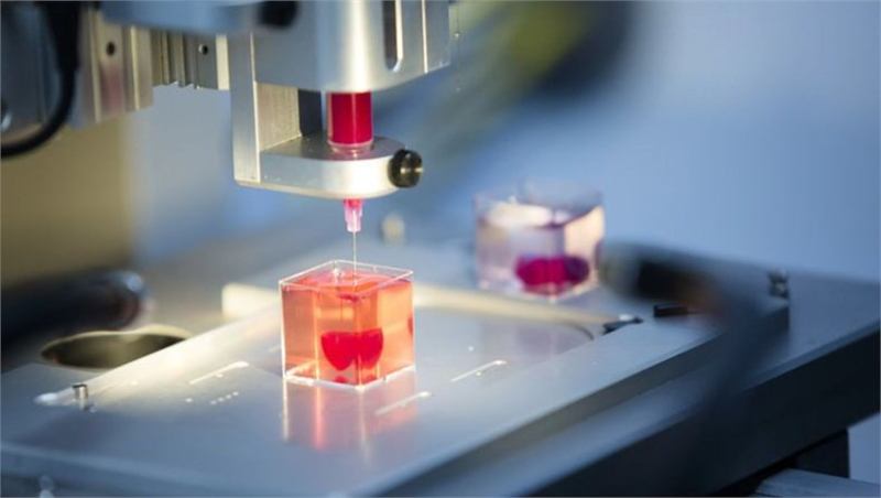
Four deposition methods dominate. Extrusion squeezes a continuous filament of bioink through a nozzle — cheap, versatile, and by far the most common. Inkjet fires individual droplets, which is fast and gentle but limited to low-viscosity inks. Laser-assisted printing uses a laser pulse to propel tiny volumes of cell suspension with no nozzle at all, giving excellent resolution and cell survival at the cost of speed and complexity. Light-based methods (stereolithography, digital light processing and volumetric printing) cure a whole layer — or in the volumetric case an entire object — at once using patterned light, which is dramatically faster.
How bioprinting works compares the four in detail, including resolution, speed and typical cell viability.
What actually happens, step by step
Imaging and modelling
The process usually starts with a CT or MRI scan of the patient, which is segmented into a 3D model. For a bone or cartilage implant the external geometry is what matters. For soft tissue, the internal architecture matters far more — where the vessels run, how dense the cells should be in each region, which cell type goes where. That model is then sliced and converted into printer instructions.
Cell expansion
A biopsy yields far too few cells, so they are cultured until there are enough — typically tens or hundreds of millions. This stage alone can take weeks and is a major reason patient-specific tissue is expensive. Cells that are grown too long begin to drift from their original identity, which puts a real ceiling on how much expansion is safe.
Printing
The cells are mixed into the bioink and loaded into the printer. Most serious constructs are printed with more than one material at once: a cell-laden bioink for the tissue itself, a stiffer polymer for structural support, and often a sacrificial ink that is printed where channels are wanted and then dissolved away afterwards, leaving hollow tubes that can be seeded with vessel-lining cells.
Cross-linking and maturation
Immediately after printing, the bioink is locked into place — by ultraviolet or visible light, by a calcium bath, by temperature, or by an enzyme. The construct then goes into a bioreactor, which perfuses it with nutrients and oxygen and often applies mechanical cues: pulsatile flow for blood vessels, stretch for muscle, compression for cartilage. Tissue that is never mechanically challenged never develops proper strength. Maturation takes days to weeks, and it is where a large share of promising constructs fail.
What bioprinting is used for right now
Transplants get the headlines, but they are not where the technology earns its keep today.
Drug testing and toxicology
This is the commercial heart of the industry. Roughly nine out of ten drug candidates that enter human trials never reach approval, and a substantial share fail on unexpected toxicity — often liver or heart toxicity that animal models did not predict, because mouse livers are not human livers. Bioprinted human liver, kidney, cardiac and tumour tissue gives pharmaceutical companies a human-relevant test system weeks before a human is ever exposed. Organovo's bioprinted liver tissue, sold from the mid-2010s, was the first widely available commercial product of this kind.
Regulatory momentum is behind this. The US FDA Modernization Act 2.0, signed at the end of 2022, removed the blanket statutory requirement for animal testing before human trials and explicitly recognised alternatives including organ chips and other cell-based models.
Cosmetics and consumer safety
The European Union has banned animal testing for cosmetics since 2013, which created immediate demand for engineered human skin. L'Oréal, Procter & Gamble and others have invested in bioprinted skin models for exactly this reason.
Implants and grafts that have reached patients
Acellular and simple cellular constructs are already in clinical use or advanced trials: patient-specific resorbable bone implants, cartilage repair, tracheal splints, electrospun dura mater substitutes, and skin grafts. In 2022 a US company implanted a 3D-printed ear built from a patient's own cartilage cells — a genuine first for a printed, cell-based implant in a human.
Surgical planning
Not bioprinting in the strict sense, but worth knowing: printing a solid replica of a patient's heart or tumour from their scan lets surgeons rehearse a difficult operation beforehand. This is routine in many hospitals and is the most widely adopted form of medical 3D printing by a wide margin.
The vascularisation problem
Every cell in your body sits within roughly 100 to 200 micrometres of a capillary — about the thickness of a sheet of paper. Beyond that distance, oxygen cannot diffuse fast enough and the cell dies. This is not a limitation of bioprinting; it is a limitation of diffusion.
The consequence is stark. You can print a sheet of skin or a corneal disc, because nothing in them is far from a surface. You cannot print anything thicker than a few millimetres without building a blood supply into it at the same time — and a human kidney contains on the order of a million filtering units served by kilometres of vessels branching down to capillaries a few micrometres across. No printer resolves that today, and even if one did, the vessels would then have to be lined with endothelial cells, made leak-proof, and connected to the patient's own circulation in surgery.
Progress is real but partial. Sacrificial inks reliably produce channels down to a few hundred micrometres. A widely cited 2019 study in Science by Bagrat Grigoryan, Jordan Miller and colleagues printed hydrogel constructs containing entangled vascular networks, including an air-sac structure that oxygenated flowing red blood cells — a striking demonstration that printed architecture can perform a real physiological function. Other groups let the tissue help: seed endothelial cells and let them self-assemble the smallest capillaries, which biology does far better than any nozzle.
Why this matters for every claim you read
When a headline says a heart has been printed, it is almost always describing either a centimetre-scale construct or a full-size acellular scaffold. Both are real achievements. Neither can be transplanted, because neither has a working blood supply. Checking for that one detail will tell you more about a bioprinting story than anything else in it.
The other hard problems
| Problem | Why it is hard |
|---|---|
| Cell sourcing | Producing hundreds of millions of the right cell type, from the right patient, reliably and without the cells losing their identity in culture. |
| Resolution versus viability | Finer nozzles give better detail but higher shear stress. Native tissue is organised at the micrometre scale; most extrusion printing works at hundreds of micrometres. |
| Maturation | A freshly printed construct is an arrangement of cells, not a tissue. Turning one into the other takes weeks of bioreactor conditioning and often does not work. |
| Scale-up and cost | A patient-specific organ is, by definition, a batch of one. Manufacturing, sterility and quality control processes built for mass production do not transfer cleanly. |
| Regulation | A printed organ is simultaneously a medical device, a biologic and a cell therapy. No regulator has a settled framework, and approval pathways are being written case by case. |
| Long-term behaviour | Implanted engineered tissue has to keep working for decades, remodel appropriately, and not form tumours. Only time produces that evidence. |
So when will printed organs arrive?
It depends entirely on what you mean by "organ", which is why published estimates vary so wildly. A sensible way to read the field:
- Flat and tubular tissues — skin, cartilage, bone, cornea, blood vessels, trachea. Already in patients or in trials. Expect steady, unglamorous expansion.
- Tissue patches — a bioprinted cardiac patch to support a damaged heart, or an islet construct for type 1 diabetes. Plausible within the next decade or so; several are in or approaching trials.
- Whole solid organs — a transplantable printed kidney, liver or heart. Researchers who work on this generally talk in terms of decades, and treat confident near-term predictions with suspicion. The vascularisation problem has to be solved first, and it has been the field's central obstacle for more than twenty years.
Watch for tissue patches rather than whole organs, and watch the drug-testing market rather than the transplant one. That is where the technology is compounding.
Keep reading
- How bioprinting worksThe four printing methods compared
- Bioinks explainedMaterials, cross-linking and trade-offs
- Printed organsWhere each organ actually stands
- GlossaryEvery term on this page, defined
- TimelineMilestones from 1988 to today
- CompaniesWho builds the printers and inks
Last reviewed . Educational information only — not medical advice.