A supply problem
In the United States alone, more than 100,000 people are on the national transplant waiting list, and the US Health Resources & Services Administration reports that 17 die each day waiting for an organ that never arrives.
Printers that build living tissue layer by layer, one cell at a time. Here is what bioprinting can actually do today, what it cannot do yet, and who is working on it.
A technology that could one day end the transplant waiting list — and that already quietly powers a good deal of drug testing.
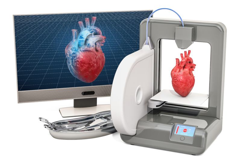
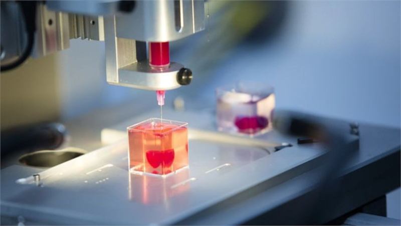
3D bioprinting works like ordinary additive manufacturing: a digital model is sliced into layers, and a machine lays those layers down one at a time. The difference is the material. Instead of molten plastic, a bioprinter deposits bioink — living cells suspended in a water-rich gel that supports them while they knit together.
The printed construct then moves to an incubator or bioreactor, where the cells reorganise, start producing their own matrix and mature into something that behaves like real tissue. Where the cells come from matters enormously: tissue grown from a patient's own cells carries far less risk of immune rejection than a donated organ.
In the United States alone, more than 100,000 people are on the national transplant waiting list, and the US Health Resources & Services Administration reports that 17 die each day waiting for an organ that never arrives.
Skin, cartilage, bone, cornea and blood-vessel grafts have reached patients or late-stage trials. Whole hearts, kidneys and livers have been printed at small scale in laboratories, but none is anywhere near transplantable.
Bioprinted liver, kidney and tumour tissue is already sold as a testing platform. Human tissue in a dish predicts toxicity better than animal models, which is why pharmaceutical companies became the industry's first paying customers.
Every tile below is a genuine laboratory or clinical result. The status label tells you how far it has actually travelled — most of this work is still pre-clinical.
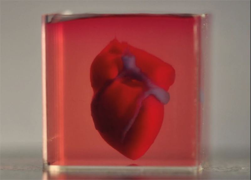
Cell-sized cardiac constructs that beat in a dish, and full-scale collagen scaffolds.
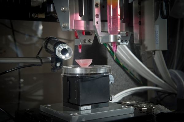
Printed nephron-like tubules and organoids used for drug toxicity testing.
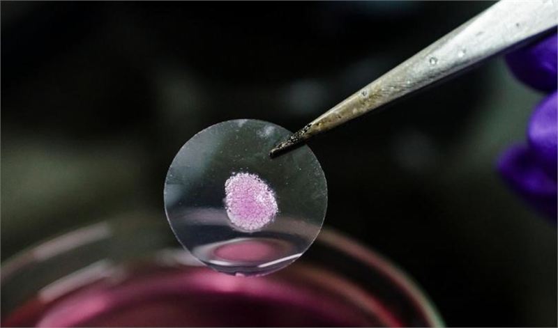
The first bioprinted tissue sold commercially, as a drug-testing model.
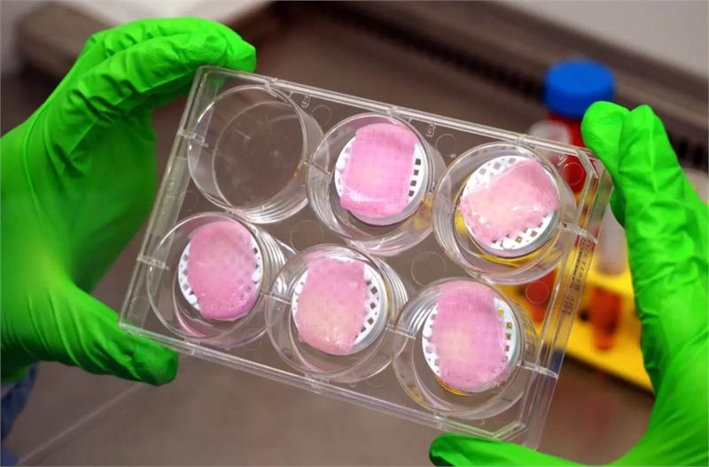
The closest to routine clinical use, including printing directly onto a wound.
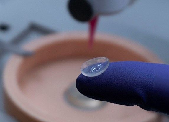
Printed in minutes from collagen and stem cells; several groups are heading for trials.
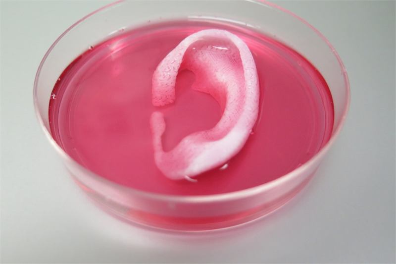
An implant grown from a patient's own cartilage cells was placed in 2022.
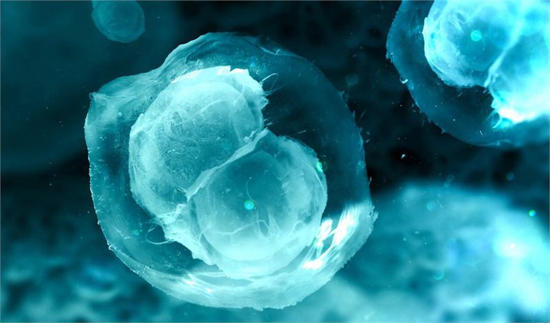
A printed scaffold seeded with follicles restored fertility in mice.
Bone, cartilage, blood vessels, trachea, nerve and pancreatic tissue.
Four stages separate a medical image from a piece of functioning tissue.
A CT or MRI scan of the patient is converted into a 3D model, then sliced into printable layers and a toolpath. Getting the internal architecture right matters more than the outer shape.
Cells — often stem cells taken from the patient — are expanded in culture and mixed into a hydrogel such as gelatin, collagen or alginate. The gel must be soft enough for cells to survive and stiff enough to hold its shape.
Extrusion, inkjet, laser-assisted or light-based printing deposits the bioink layer by layer, often alongside a sacrificial material that is later washed out to leave open channels for blood vessels.
The construct goes into a bioreactor that supplies nutrients, oxygen and mechanical stimulation. This maturation stage takes days to weeks and is where most attempts fail.
Twenty-four organisations building printers, bioinks and tissue products. Logos link to the company's own site.
























Recent coverage of bioprinting and tissue engineering from across the web.
No stories are available right now. Please check back soon.
The news feed could not be loaded. Please try again later.
Simple printed tissues have — skin grafts, cartilage, bone and a printed ear implant placed in 2022. No solid internal organ such as a heart, kidney or liver has been printed and transplanted. Those remain laboratory work.
Thickness. Cells more than roughly 200 micrometres from a blood supply starve, so any tissue thicker than a sheet of paper needs its own vascular network. Skin is thin and flat; a kidney contains around a million filtering units fed by kilometres of vessels. Building that plumbing is the field's central unsolved problem.
Simple flat and tubular tissues are already in trials and should broaden over the next decade. Researchers in the field generally place transplantable solid organs decades away, and treat any shorter estimate with caution.
That is the main reason to print at all. If the tissue is built from the patient's own cells it is genetically their own, so the lifelong immunosuppressant drugs that transplant recipients depend on may not be needed.
Corrections, missing companies, research tips and partnership enquiries are all welcome.