In April 2019, photographs of a small red heart floating in a dish of pink medium went round the world. The work was real and the achievement significant. It was also, as the researchers themselves said clearly at the time, nowhere near a transplant. Here is what was actually built, and what still has to happen.
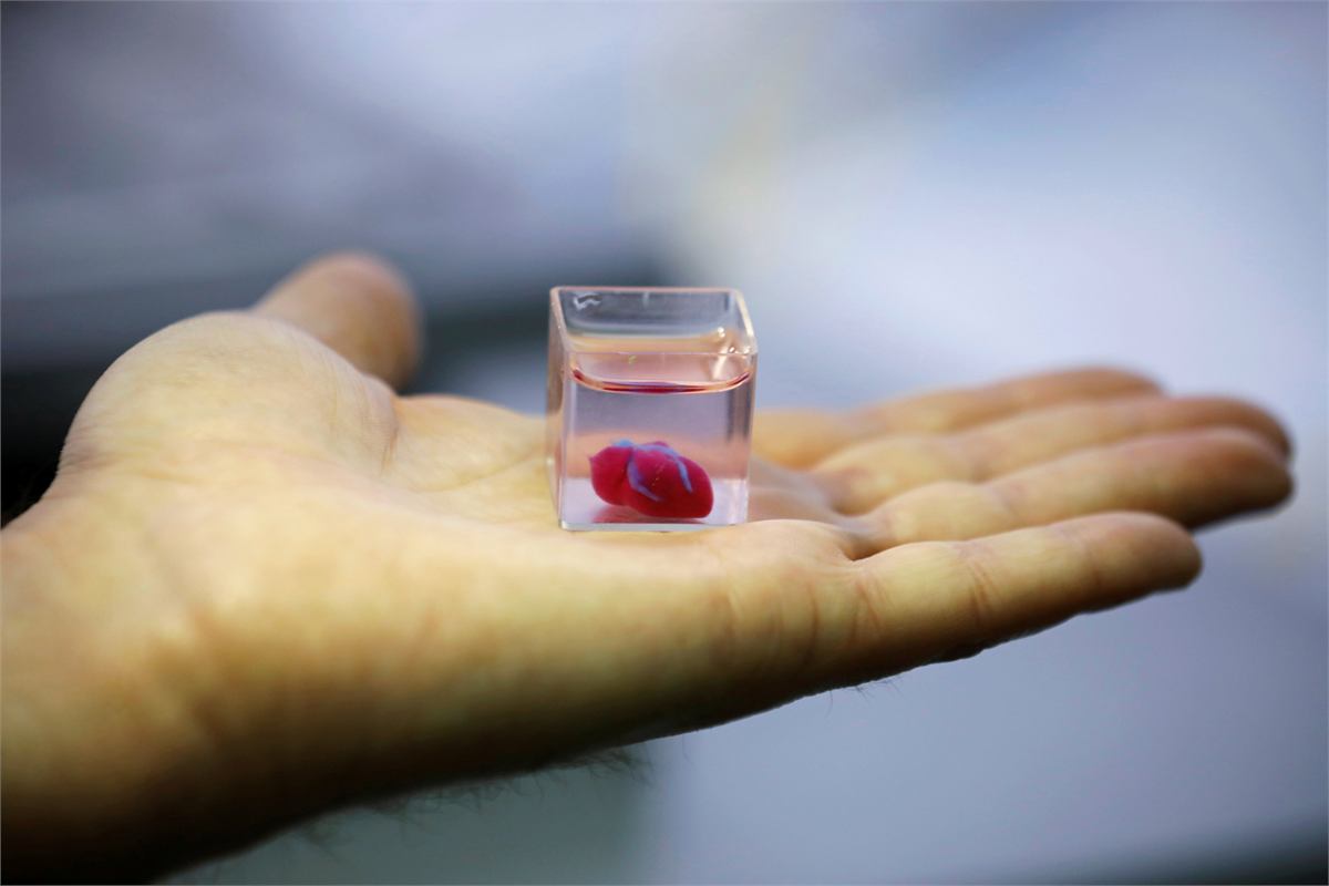
The 2019 Tel Aviv heart
A team led by Tal Dvir at Tel Aviv University reported the first 3D-printed heart made from a patient's own cells and materials, published in Advanced Science. The approach was genuinely novel in one respect that matters more than the shape.
They took a biopsy of omentum — fatty tissue from the abdomen — and separated it into two parts. The cells were reprogrammed into induced pluripotent stem cells and then differentiated into cardiac muscle cells and endothelial cells. The remaining extracellular matrix was processed into a personalised hydrogel. The result was a bioink in which both the cells and the supporting material came from the same patient, which in principle eliminates rejection entirely.
The printed construct was about 2.5 centimetres across — roughly rabbit-heart sized — with chambers and a printed vascular network. The cells contracted. What they did not do was contract together: the construct had no coordinated pumping action, because the cardiomyocytes had not matured enough to conduct an electrical signal across the whole tissue in synchrony.
What it proved, precisely
That a personalised bioink made entirely from one patient's own tissue can be printed into a complex, chambered, vascularised structure containing living heart cells. That is a substantial result. It is not a working heart, and Dvir's group said so at the time.
The FRESH collagen scaffold
Later in 2019, Adam Feinberg's laboratory at Carnegie Mellon published in Science a different kind of milestone: a full-size human heart printed from collagen.
Collagen is the material cardiac tissue is actually built from, and it is notoriously unprintable — it gels slowly and collapses under its own weight in air. The group's FRESH technique (Freeform Reversible Embedding of Suspended Hydrogels) solves this by printing inside a bath of gelatin microparticles that supports each layer until it sets, then melts away at body temperature.
Using it, they printed anatomically accurate structures at multiple scales, including capillary-sized features, functioning heart valves and a full-size neonatal heart. They also printed contracting ventricle models with real cardiomyocytes.
The trade-off runs the other way from Tel Aviv. Here the anatomy was full scale and the material ideal, but the full-size heart was a scaffold rather than a living organ — there is no practical way to supply the hundreds of billions of cells a real human heart contains.
Cardiac patches: the realistic target
Ask cardiac tissue engineers what they expect to reach patients first and almost none of them says a whole heart. They say a patch.
After a heart attack, part of the muscle dies and is replaced by scar tissue that does not contract. A bioprinted cardiac patch — a sheet of aligned, beating cardiomyocytes with its own microvasculature, sutured or glued over the damaged region — would not replace the heart, only supplement it. That is a far smaller problem: a patch is thin enough to survive on diffusion for long enough to connect to the host's blood supply.
Several such constructs, printed and otherwise engineered, have moved into early-stage human trials. Related work using cardiomyocyte sheets derived from iPS cells has been carried out in Japan. This is where the near-term clinical progress will come from.
Why a heart is so hard
- Cell number. An adult human heart contains on the order of two to three billion cardiomyocytes among several billion cells in total. Producing that many mature, patient-derived cardiomyocytes is beyond current manufacturing.
- Electrical coupling. A heart is not a bag of contracting cells; it is a conduction system. Every cell must be electrically coupled to its neighbours so that a wavefront sweeps through the muscle in the correct sequence. Poorly coupled printed tissue does not just fail to pump — it can generate dangerous arrhythmias.
- Maturity. Cardiomyocytes made from stem cells tend to resemble foetal cells: weaker, less organised, electrically immature. Getting them to adult-like function requires prolonged mechanical and electrical conditioning and remains unreliable.
- Mechanical endurance. A heart beats roughly 100,000 times a day, every day, for decades. Engineered tissue has to survive that fatigue loading.
- Vascular density. Cardiac muscle is among the most metabolically demanding tissue in the body, with capillaries on the order of one per muscle cell. Printing that density is not currently possible.
When might a printed heart be transplanted?
Nobody serious gives a date. What can be said is what has to happen first, roughly in order: reliable manufacturing of mature cardiomyocytes at scale; printed vascular networks that reach capillary dimensions and stay open; bioreactor conditioning that produces adult-like contractile function; demonstration of years of stable function in large animals; and a regulatory pathway that does not yet exist.
Each of those is a multi-year programme, and several are unsolved research problems rather than engineering tasks. Researchers in the field typically speak in decades. Meanwhile, printed cardiac tissue is already doing useful work in drug testing, where a beating human cardiac construct catches the cardiotoxicity that has forced more than one approved drug off the market.
Related
- All printed organsThe full status report
- KidneyThe hardest organ of all
- FRESH and support bathsHow collagen became printable
- Timeline2019 in context
Last reviewed . Educational information only — not medical advice.